What it is
The retina lines the inside of the eye like wallpaper in a room. It receives light and sends the image to the brain. To work, it has to stay attached to the wall, which is what nourishes it.
In the most common type, it all starts with a tear. Fluid seeps through that hole behind the retina and lifts it, just as water peels paper off a wall. The detached part stops seeing.
What matters most is the macula, the centre of the retina, which you use to read and recognise faces. If the detachment has not reached it yet, operating in time usually saves central vision. If it has, vision only recovers in part.
There are other, less common types, such as the one seen in advanced diabetic retinopathy, where membranes pull on the retina. This guide covers the usual one, which starts from a tear.
How it shows
It does not hurt. It warns you in other ways:
- Flashes, like lightning or camera flashes at the side of your vision.
- Many new floaters all at once, or a shower of black dots.
- A shadow or curtain coming in from one side and moving towards the centre over hours or days.
- Loss of central vision, once the detachment reaches the macula.
A spreading shadow is an emergency. Call the same day. Flashes and new floaters also need checking soon, because they are often the warning of a tear that can still be sealed with laser. I explain this in the guide on floaters, flashes and retinal tears.
Who is most at risk
- Short-sighted people, especially if highly so.
- People who have had cataract surgery.
- People who have had a detachment in the other eye, or whose parent or sibling has had one.
- People who have had a hard blow to the eye.
- People with weak areas at the edge of the retina, such as lattice degeneration.
It is more common from the age of 50, when the gel that fills the eye separates from the retina and sometimes tears it.
Why it is urgent
A detachment does not heal on its own and almost always progresses. Without surgery it usually ends up involving the whole retina, and the sight in that eye is lost.
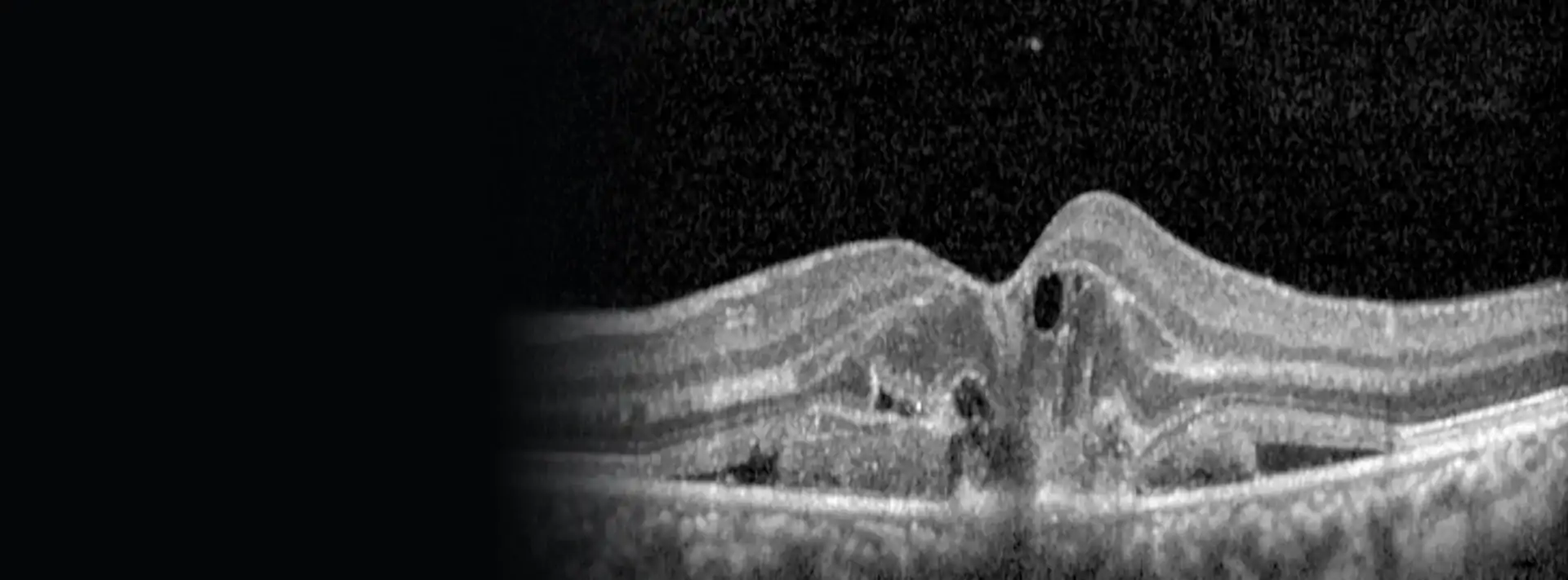
I dilate the pupil and examine the whole retina to find the tears and see how far the detachment extends. The OCT tells me whether the macula is still in place, and that sets the pace:
- Macula still attached: surgery as soon as possible, within days, to get there before it comes away.
- Macula already detached: surgery is still done promptly, but a few days change the outcome less.
Until the operation I will tell you which position to rest in. It helps keep the fluid from moving towards the centre.
Surgery
Vitrectomy
This is the operation I do in most cases. Through three tiny incisions, of 23, 25 or 27 gauge, I remove the vitreous, the gel that pulls on the retina. I then drain the fluid from behind it so that it rests against the wall again, seal the tears with laser and fill the eye with a gas that holds it in place while it heals.
It is done under local anaesthetic: I numb the eye with an injection beside it (retrobulbar anaesthesia) and you feel no pain. The incisions usually close without stitches.
Reattaching the retina during vitrectomy
Once the vitreous is removed, the detached retina settles back into place.
Argon laser in retinal detachment
Laser sealing around the tear so the retina stays fixed.
Scleral buckle, in complicated cases
Sometimes the retina forms abnormal scar tissue that pulls on it and makes it detach again. This is called PVR. When it appears, or when there has already been a re-detachment, I add a scleral buckle: a thin band placed around the outside of the eye that helps keep the retina in place.
In the most complex cases, silicone oil is left inside the eye instead of gas. It supports the retina for longer and is removed in a second operation.
Complex cases, on video
Four of my own operations on difficult detachments. These are real microscope images, and they only play if you press play.
Tractional detachment in diabetes
The fibrovascular proliferations caused by poorly controlled diabetes are stained with blue dye and progressively peeled away to reattach the retina.
Re-detachment from scar tissue (PVR)
Removing the membranes that had detached the retina again, in an eye filled with silicone oil.
Detachment after a retinal infection
The membranes are released so the retina can settle back into place.
Surgery guided by live OCT
The microscope's OCT shows the retina in cross-section while I operate. Here it guides the removal of a tiny bubble left under the centre of the retina.
If you also have a cataract
I can operate on the cataract and the retina in the same procedure. I suggest it when the cataract is already bothering you or stops me seeing the retina clearly during surgery.
If there is only a tear
When a tear is found before the retina comes away, no operating theatre is needed. I seal it with argon laser in the clinic, in a few minutes.
After the operation
Posture
The gas bubble floats. For it to press exactly where the tear was, you will need to keep your head in a particular position for a few days. Which one, and for how long, depends on where the tear is, and I explain it before the operation so you can get organised at home.
While there is gas in your eye
Vision in that eye will be very blurred. Depending on the gas, the bubble takes from a couple of weeks to a couple of months to be absorbed. You will see it as a line that moves and drops lower as it gets smaller. While it lasts:
- Do not fly or go up into the mountains or over high passes: the gas expands with altitude and the pressure in the eye can rise dangerously.
- If you need another operation under general anaesthetic, say that you have gas in your eye. One anaesthetic (nitrous oxide) must not be used.
- Use the drops I prescribe and avoid heavy exertion.
Call straight away if, after the operation, you notice:
severe pain that does not settle, an eye that is getting redder, vision getting worse instead of better, or the shadow coming back.
Results
Today the retina can be reattached in more than 9 out of 10 detachments. Some cases need more than one operation, nearly always because of the abnormal scar tissue mentioned above.
A retina back in place does not mean vision returns to what it was. That depends mostly on whether the macula came away and for how long. If surgery was in time, good vision is usually kept. If the macula detached, improvement is slow, over months, and the image may stay slightly distorted.
- If you have not had cataract surgery, a cataract often develops in the months or years after a vitrectomy. It can be treated.
- Your other eye is at higher risk than someone else's. I always check it, and if I find a tear or a weak area I treat it with laser.
- If flashes, floaters or the shadow come back, in either eye, call without waiting for your check-up.
Seeing flashes, many new floaters or a spreading shadow?
Call today. If the retina is coming away, every day counts.