What it is
The macula is the centre of the retina, the part you use to read and recognise faces. Here we are talking about two different problems that cause very similar symptoms, and that often go together.
Epiretinal membrane
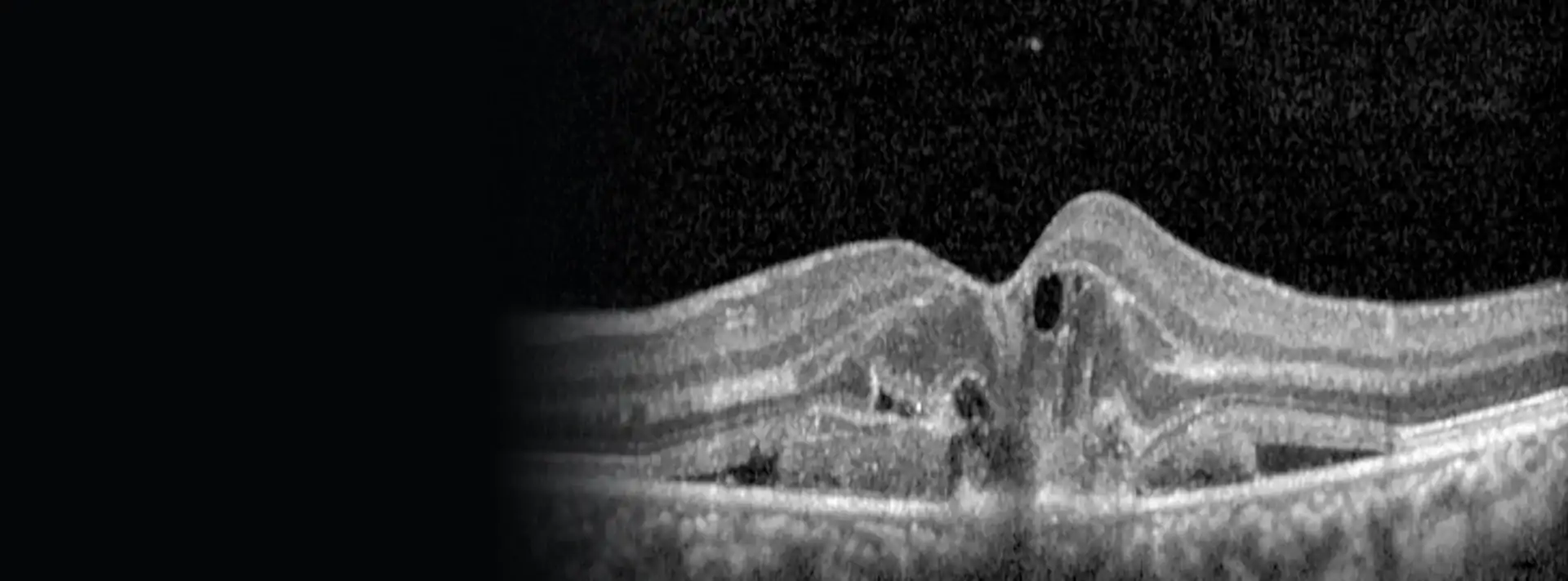
This is a very thin layer, like cling film, that grows on the surface of the retina. If it stays still, it barely causes any trouble. If it contracts, it pulls on the macula and wrinkles it, just as tissue paper wrinkles when it shrinks.
Vitreomacular traction
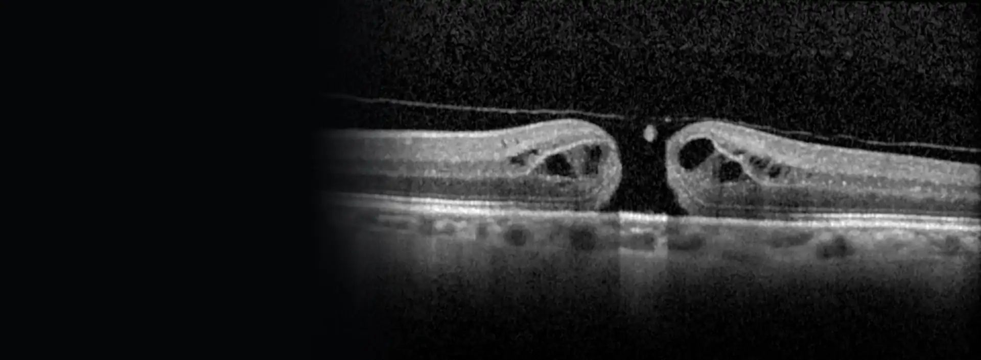
Inside the eye there is a gel, the vitreous. With age it comes away from the retina. Sometimes it separates everywhere except the centre, and that point which is still attached pulls the macula forwards and distorts it.
When the vitreous is still attached to the macula but does not distort it, it is called vitreomacular adhesion. This is a normal stage in the ageing of the vitreous, it causes no symptoms and is not treated.
The word "idiopathic" that you will see in some reports just means there is no specific cause. That is the most common situation.
Symptoms
Many people have a membrane and don't know it: it is found at a check-up. When it does cause symptoms, the typical ones are:
- Straight lines looking bent: a door frame, floor tiles, lines of text.
- Blurred or less sharp vision in that eye.
- One eye seeing things at a different size from the other, making it hard for both eyes to work together.
- Double vision in some cases.
- Difficulty reading or driving. Some people end up closing the affected eye to read better.
It usually progresses slowly, over months or years. With vitreomacular traction the symptoms can come on more suddenly.
Book an appointment soon if a patch appears in the centre of your vision, if lines suddenly look bent or if your sight drops quickly. Sometimes the traction ends up opening a macular hole, and in that case it is best to act without waiting months. Let me know as well if you notice lots of new floaters or a shadow on one side of your vision.
Who gets it
It is a common problem, and more so the older you are. An OCT scan finds it in a significant proportion of older people, although most have very mild forms. In some people it appears in both eyes.
As well as age, it is more common in people who have had:
- A vitreous detachment, which is when the gel comes away from the retina.
- Retinal tears or a retinal detachment.
- Inflammation inside the eye (uveitis).
- A retinal vein occlusion or diabetic retinopathy.
- Cataract surgery or other eye operations.
Other factors, such as short-sightedness or smoking, have been studied, but it is not clear that they play a part. There is nothing specific you can do to prevent it.
How it is diagnosed
I examine the back of the eye with the pupil dilated, including the outer edge of the retina to rule out tears. And I do an OCT scan, which is the key test: in a few seconds, without touching the eye, it shows me a cross-section of the retina. It shows the membrane, whether the vitreous is pulling, how much the macula has thickened and whether there is fluid or a hole.
The OCT also lets you see it for yourself. Putting your scan next to a normal retina helps a lot in understanding what is happening and why I suggest one option or another.
If I suspect the membrane is caused by another retinal condition, I may add other imaging tests.
Monitoring without surgery
Here is the reassuring news: most membranes do not change much over the years. In population studies, only a little under a third got worse in five years, and some even improved. Waiting until there are symptoms does not worsen the result of an operation later on.
With vitreomacular traction, especially when the attached area is small, the vitreous lets go by itself in quite a few cases, roughly a third within one or two years. When that happens, vision usually improves.
In the meantime:
- Check-ups with OCT to compare with the previous one.
- Check each eye separately from time to time, covering the other one. The Amsler grid or the lines of a door frame will do.
- There are no drops or tablets that get rid of a membrane.
What about the injection to release the vitreous?
There was an injectable drug, ocriplasmin, designed to release the traction. In the European Union it was withdrawn from the market in 2023 by the manufacturer for commercial reasons, so it is not available in Spain today. Injecting a gas bubble to detach the vitreous has also been tried, but one study was stopped early because of retinal detachments. It is not a routine option.
Surgery
When to operate
It mostly comes down to how much it bothers you. If the distortion or blurred vision makes it hard to read, drive or get on with daily life, surgery makes sense. If you barely notice it, the sensible thing is usually to monitor it.
It is not an emergency. Operating early may give slightly better vision in the long run, but we are talking about months, not days. In vitreomacular traction with a wide attached area, fluid under the retina or vision that is already low, it is less likely to resolve on its own and it is usually better not to wait as long.
What it involves
The operation is called a vitrectomy. I make three tiny openings in the wall of the eye with 23G, 25G or 27G instruments, so fine that they usually don't need stitches. I remove the vitreous, release any traction and, with very fine forceps, peel the membrane off the surface of the retina.
I often also remove the internal limiting membrane, a clear layer of the retina that acts as scaffolding for the membrane. Removing it lowers the chance of the membrane growing back. To see it I use a dye that stains it during the operation.
It is done under local anaesthetic with a retrobulbar injection, behind the eye: the eye goes numb and stays still, and you feel no pain. You can see what it looks like in the video of vitrectomy with ILM peeling.
If you also have a cataract
After a vitrectomy, most people who still have their natural lens develop a cataract in the following months or years. If you already have a cataract, it can be operated on before, at the same time or afterwards. The results are similar, so we decide based on your case.
Results and risks
Overall, about 8 in 10 people who have the operation clearly gain vision. The distortion usually improves a lot, and sometimes disappears. Some people don't gain many letters on the eye chart and are still happy, because lines have stopped looking bent.
It is worth having realistic expectations: improvement is slow, takes months, and is not always complete. Between 1 and 2 in 10 patients stay the same or are slightly worse. If the deeper layers of the retina look well preserved on OCT, the outlook is better.
The risks I explain to you before surgery:
- Cataract: the most common consequence, almost certain over time if you haven't already had cataract surgery.
- Retinal tear or detachment: uncommon with modern surgery, around 1 in 100.
- The membrane coming back, less likely if the internal limiting membrane is removed.
- Changes in eye pressure in the first few days, especially if you have glaucoma.
- Serious infection inside the eye (endophthalmitis): very rare, fewer than 1 in 2000 operations.
After your operation
I'll see you the next day and again after one or two weeks, sooner if needed. I'll give you some drops and explain how to put them in.
If I leave a gas bubble in the eye at the end, your vision will be blurred until it is absorbed and you will need to take precautions while it lasts: no flying, no going up into the mountains, and telling the team you have it if you need a general anaesthetic. I'll give you the specific instructions for your case.
If after the operation you still see double or find it hard to use both eyes together, there is specific help available from binocular vision specialists.
Call straight away if after the operation you notice pain that keeps getting worse, your vision gets worse, lots of new floaters appear, or a shadow or curtain covers part of your vision. These can be signs of high pressure, infection or retinal detachment, and in all of them acting early changes the outcome.
Are straight lines looking bent, or have you been told about a membrane on your retina?
Book an appointment. With an OCT scan at the clinic I'll show you how your macula looks and we'll decide together whether to monitor it or operate.