What a vitreous detachment is
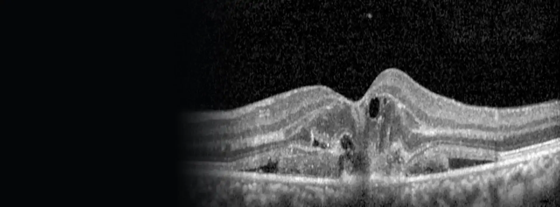
The inside of the eye is filled with a clear gel, the vitreous. When we are young it is firm and attached to the retina, the layer at the back of the eye that picks up the image. Over the years it becomes more liquid, shrinks and eventually comes away from it.
That separation is a posterior vitreous detachment. It is not a disease: it happens to nearly all of us, usually between the ages of 45 and 65. In short-sighted people, after a blow to the eye or after cataract surgery it can come earlier.
The name sounds alarming because it is similar to "retinal detachment", but it is not the same thing. What comes away is the gel, not the retina.
Floaters and flashes
When the vitreous comes away, you will usually notice one or more of these:
- Floaters: dots, threads, cobwebs or a ring that move when you move your eye and are easier to see against a white wall or the sky.
- Flashes: sparks or streaks of light, often off to one side, that are more noticeable in the dark. They appear when the gel pulls on the retina as it separates.
- Slightly hazier vision, as if you were looking through water.
Over weeks or months the flashes fade and the brain learns to ignore the floaters. Many do not go away completely, but they stop bothering you.
If you get new floaters, flashes, or a shadow or curtain covering part of your vision, call that same day. The symptoms alone cannot tell us whether there is a tear: the pupil has to be dilated and the retina examined. And if the shadow is spreading, the retina may already be coming away.
When it gets complicated: retinal tears and detachment
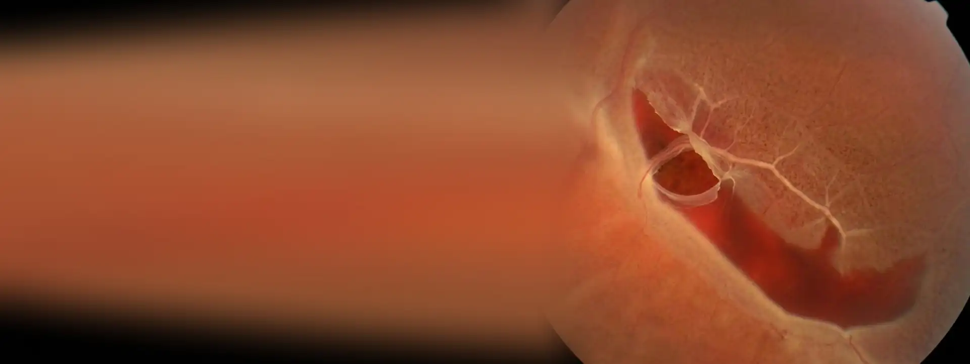
In some places the vitreous is very firmly stuck to the retina. If it pulls hard as it separates, it can tear it. This happens in around 1 in 15 people with a recent vitreous detachment, and it is more likely if there is blood inside the eye (a sudden shower of black dots).
A retinal tear is a hole through which fluid can seep underneath the retina and lift it. That is then a retinal detachment: the part that has come away stops seeing, and if it reaches the centre you lose your reading vision.
The good news is that there is time to act. Without treatment, at least half of tears that cause symptoms end up detaching the retina. Treated early with laser, the risk drops to less than 1 in 20.
Sometimes the tear appears days or weeks after the first floaters. So even if your first check-up is fine, it is worth coming back if anything changes.
Who is at higher risk
A retinal tear or detachment is more likely in people with:
- Short-sightedness (myopia), especially if it is high. More than half of detachments not caused by an injury happen in short-sighted eyes. Laser surgery for short-sightedness corrects the prescription, but not the risk to the retina.
- Lattice degeneration in the outer part of the retina (I explain it further down).
- Cataract surgery or other surgery inside the eye, and also the YAG laser used after cataract surgery.
- A previous retinal detachment in the other eye, or in a parent, brother, sister or child.
- Blows to the eye, even years ago.
- Some genetic conditions, such as Stickler syndrome, and certain eye drops that make the pupil smaller (for example pilocarpine, which is now also advertised for age-related reading difficulty).
If you have any of these factors, there is no need to live in worry. You just need to know the warning signs and not skip your check-ups.
Lattice degeneration
This is an area of thinner retina towards the edge, with the vitreous firmly stuck to its borders. Between 6 and 8 in 100 people have it, many without knowing, and it is more common in short-sighted people. It causes no symptoms: it is found during an examination of the back of the eye.
It sounds more worrying than it usually is. Most people with lattice degeneration will never have a detachment, even if there are small round holes within it. That is why, in general, it is not treated as a precaution: it is monitored.
When do I treat it?
What I check in clinic
I put in some drops to dilate your pupil and examine the whole retina, including the outermost part, which is where tears are usually found. Sometimes I press gently on the eyelid from the outside to see right to the edge. It is uncomfortable, but it does not hurt.
The OCT lets me see how the vitreous is separating in the centre of the retina, and retinal photography keeps a picture to compare at later visits. If there is so much blood inside the eye that the back cannot be seen, an ultrasound scan is needed to look for tears behind it.
The dilating drops blur your vision for a few hours. Come with someone and do not drive home.
Treatment
Vitreous detachment without a tear
It needs no treatment. Floaters cannot be removed with drops or tablets. What matters is knowing which symptoms should bring you back.
If after several months the floaters are still so troublesome that they stop you living normally, in specific cases a vitrectomy to remove them can be considered. Laser to break them up still has little evidence in its favour.
Retinal tear: laser
It is treated with argon laser, in the clinic, with no need for an operating theatre. With a lens resting on the eye and anaesthetic drops, I apply small laser spots around the tear. They leave a scar that "welds" the retina in place and acts as a barrier to stop fluid getting through. You will see flashes of light and, at most, some discomfort.
The scar takes a few weeks to become firm. In some cases, especially if the tear is very far out or the view is hazy, freezing (cryotherapy) is used instead of laser.
Retinal detachment: surgery
If the retina has already come away, laser is not enough and an operation is needed. Depending on the case I do a vitrectomy (I remove the vitreous from inside the eye and put the retina back in place). In complicated cases, when PVR develops (abnormal scarring of the retina that pulls on it and makes it detach again) or when there has already been a new detachment, I add a scleral buckle: a thin band placed on the outside of the eye that helps hold the retina in place. Today the retina can be reattached in more than 95 in 100 uncomplicated detachments, although sometimes more than one operation is needed.
The sooner the operation, the better the vision, especially if it is done before the centre comes away. In the surgery section you can watch videos of argon laser and of a retinal reattachment with vitrectomy. The full explanation is in the retinal detachment guide.
Check-ups and warning signs
If you have a vitreous detachment with symptoms and your retina is fine, I see you again after about 4 weeks. Sooner if there was some blood. After treating a tear, check-ups are closer together at first and then spaced out to once a year.
With lattice degeneration or small holes and no symptoms, a check-up once a year or every one or two years is enough, depending on what I find.
Even when a tear has been treated well, new ones can appear over time, especially in eyes that have had cataract surgery. So the rule is simple and applies for life:
- New floaters, or many more than you had.
- Flashes of light you did not have before.
- A shadow, veil or curtain over part of your vision.
- A drop in vision with no explanation.
If you notice any of these, call that same day. Do not wait to see if it goes away.
Have floaters or flashes suddenly appeared?
Call today. With your pupil dilated I check the whole retina, including the edges, and if there is a tear I treat it with laser as soon as possible.