What it is
The macula is a tiny area in the centre of the retina. It gives you detailed vision: reading, sewing, driving, seeing the face of the person in front of you. In AMD this area gets damaged over the years.
It usually appears after 50. It affects central vision and spares side vision, so even when it is advanced, it is very rare for someone to go completely blind. People can find their way around the house and walk on their own, but they struggle to read or make out faces.
Dry and wet: not the same thing
Dry AMD
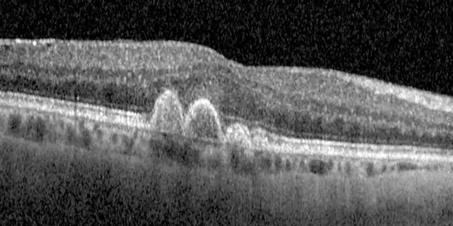
This is the most common: about 8 in 10 people with AMD have it. It progresses slowly, over years. Yellowish deposits called drusen can be seen in the retina. Its advanced form is called geographic atrophy.
Wet AMD
Less common, but it causes most cases of severe sight loss. Abnormal blood vessels grow under the retina and leak fluid or blood, and vision can get worse within weeks. This is the form that has effective treatment.
Dry AMD goes through stages: early (small drusen, almost no risk), intermediate (medium or large drusen, with a higher risk of progressing) and advanced. Dry AMD can turn into wet AMD at any time. That is why check-ups matter.
Symptoms
At first you may not notice anything. When it does show, the typical signs are:
- Straight lines that look bent or wavy: door frames, floor tiles, the line of text in a book.
- A dark or blurred patch in the centre of what you are looking at.
- More difficulty reading, especially in dim light.
- Taking longer to adjust when you go from a bright place to a dark one.
- Colours looking less vivid.
If lines suddenly look bent or a patch appears, book an appointment as soon as possible. It could be the wet form, and with that form every day counts: treated in time, much more vision is kept.
Risk factors
- Age, the main factor.
- Smoking, the most important of the ones you can change. It multiplies the risk, and so does other people's smoke. Stopping helps: over the years, an ex-smoker's risk gets closer to that of someone who never smoked.
- Having relatives with AMD. Genes are involved, but routine genetic testing is not recommended today: it does not change the treatment.
- Diet. A Mediterranean diet (vegetables, fruit, pulses, fish) is linked to a lower risk and to the disease progressing more slowly.
What about aspirin? If your doctor has prescribed it, keep taking it. The studies do not justify stopping it for the sake of your eyes.
How it is diagnosed
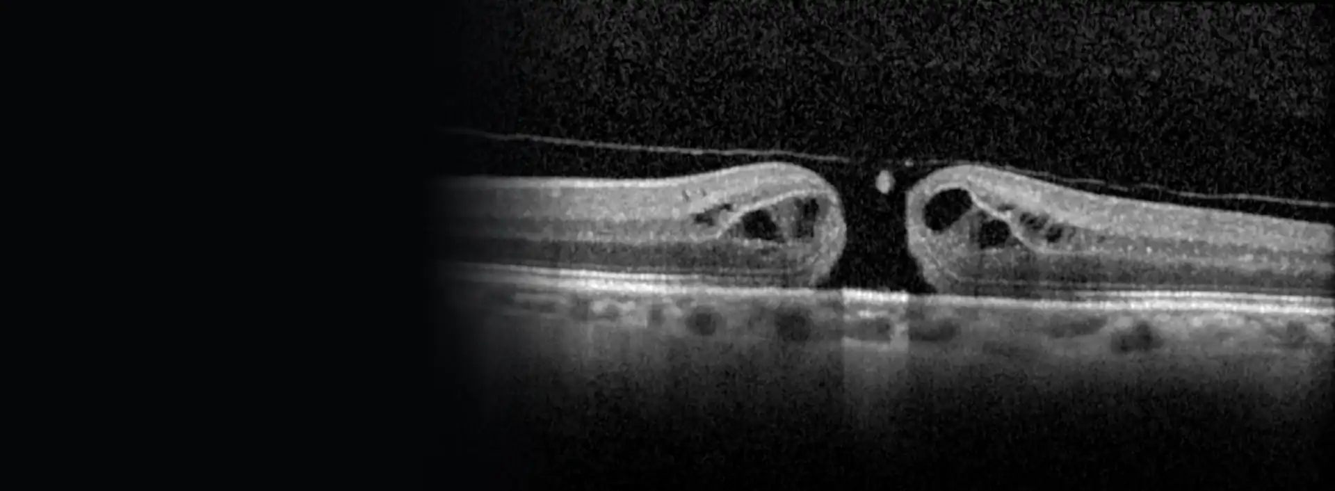
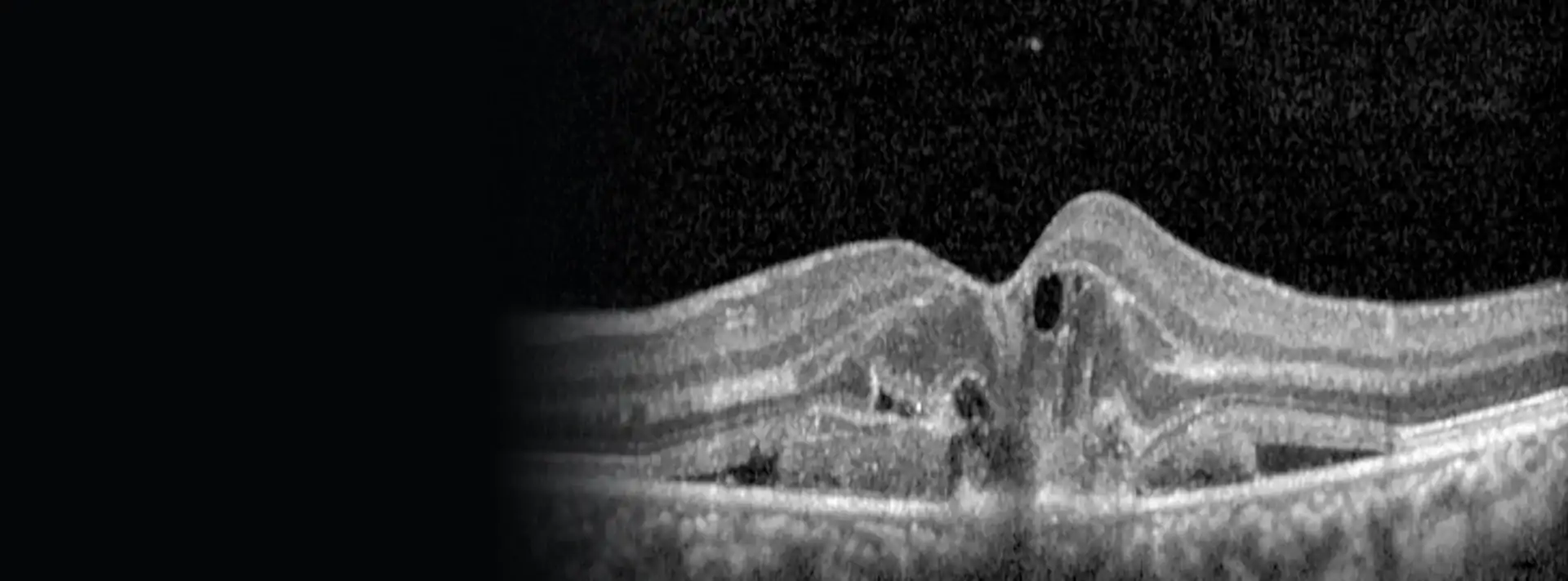
With an examination of the back of the eye and, above all, with OCT, which shows me the retina layer by layer in a few seconds without touching the eye. It picks up fluid that is not yet causing symptoms, and lets me compare each check-up with the previous one.
Depending on the case I add a retinal photograph (a picture of the back of the eye) or other imaging tests to confirm whether there are abnormal vessels.
Treatment
Early AMD
No treatment has been shown to slow it down. What works is keeping watch: check-ups, the Amsler test at home and stopping smoking.
Intermediate AMD, or advanced in one eye
Here specific supplements do help, with the formula from the AREDS studies: vitamin C, vitamin E, lutein, zeaxanthin, zinc and copper. At these stages they reduce the risk of moving on to the advanced form by around a quarter.
They are not for everyone: they have shown no benefit in the early stage, nor for preventing it in healthy relatives. Ask me before taking them.
Wet AMD
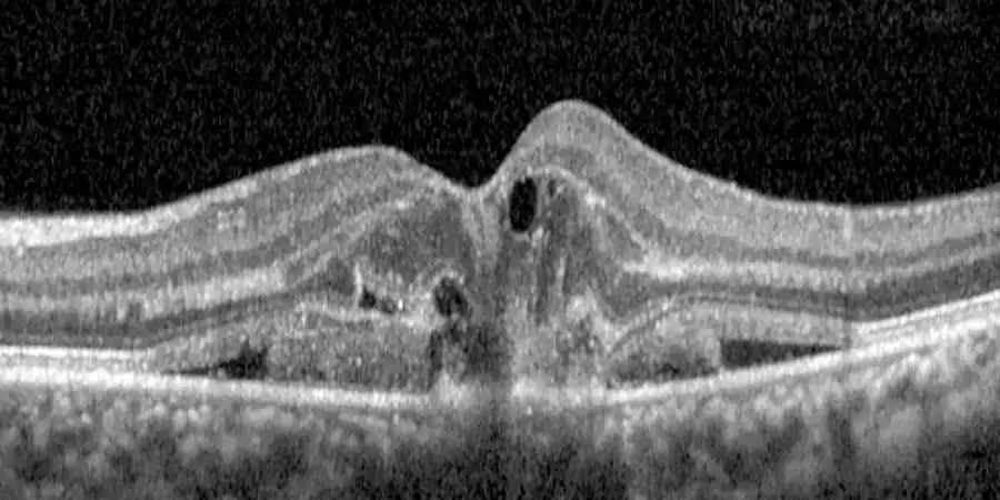
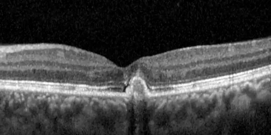
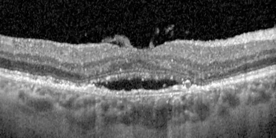
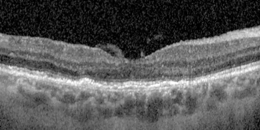
The first-choice treatment is intravitreal injections of anti-angiogenic drugs (anti-VEGF drugs). They hold back the abnormal vessels and dry up the fluid. They are given with anaesthetic drops, in a few minutes, and usually all you feel is pressure.
They control the disease but do not cure it: they have to be repeated. At first they are closer together and, if the retina responds, they are spaced out. With the newer drugs, many patients can go three or four months between injections.
Case 1
 Before
 After
AfterCase 2
 Before
 After
AfterCase 3
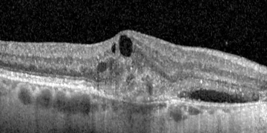 Before
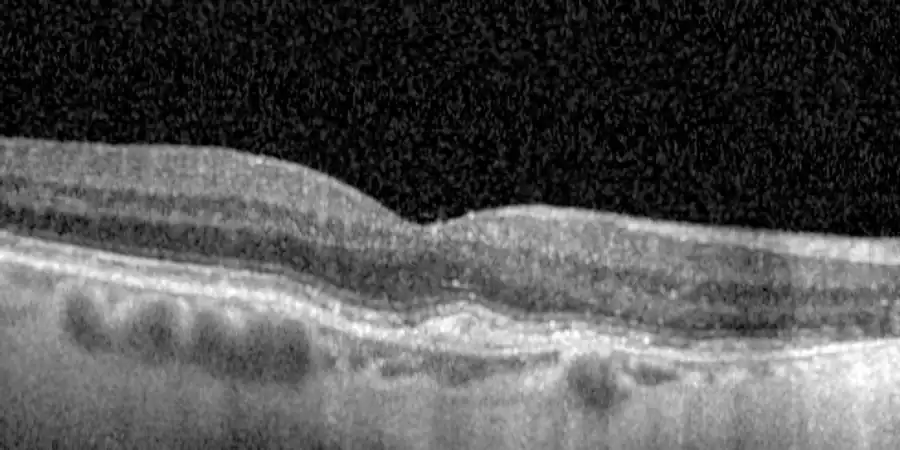 After
AfterAfter an injection, call straight away if you notice:
Geographic atrophy
In the United States two injectable drugs have been approved that slow the growth of the lesions, although in the trials they did not improve vision. In Europe the European Medicines Agency has not authorised them, so they are not available in Spain today. Management is based on monitoring, supplements when they are indicated, and low vision aids.
The Amsler test, at home
It is the simplest way to keep an eye on yourself between check-ups. Do it once a week, in good light:
- 1Put on your reading glasses, if you use them.
- 2Sit about 30–35 cm from the screen.
- 3Cover one eye with your hand.
- 4Look steadily at the dot in the centre, without moving your gaze.
- 5Notice whether any line looks bent or blurred, or whether any part is missing.
- 6Repeat with the other eye.
If you notice anything new compared with the week before, call me. Don't wait for your next check-up.
Living with AMD
- Using your eyes does not wear them out. Read, sew or look at your phone without worrying: it will not make you worse.
- Good lighting, magnifiers and electronic low vision aids let you keep reading when the macula is already affected. I can advise you on which ones might suit you.
- With poor central vision the risk of falls goes up. Take care with stairs, rugs and kerbs.
- Some people with significant sight loss see shapes or pictures that are not there. This is called Charles Bonnet syndrome. It is common and has nothing to do with losing your mind. Tell me about it at your appointment without worrying.
- If your mood suffers, say so. It is common and help is available.
Have you been told you have AMD, or are straight lines looking bent?
Call and I'll give you an appointment. With an OCT scan at the same visit we'll know where you stand.