What it is
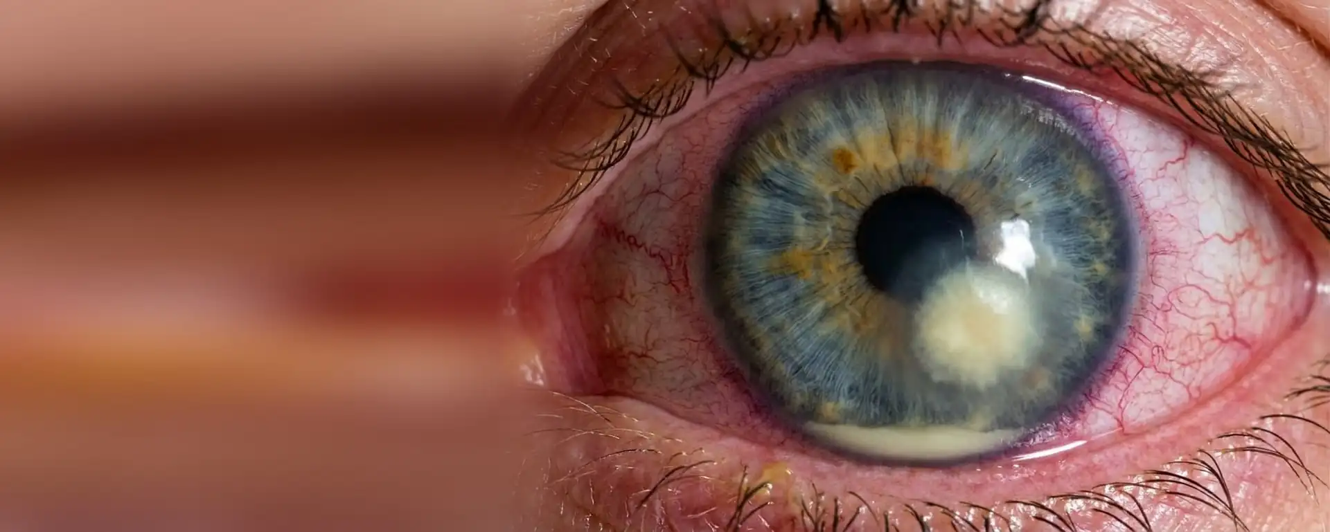
The cornea is the clear layer that covers the coloured part of the eye. On the outside it is protected by a very thin skin, the epithelium. If that skin is damaged, even by the slightest scratch, bacteria can get in and cause an infection. That is bacterial keratitis, and when it leaves an open sore on the cornea we call it a corneal ulcer.
It is not conjunctivitis. It is more serious. Some bacteria move very fast and can leave a white scar that blocks your vision, or even make a hole in the cornea. Treated in time, most cases heal well.
Symptoms
- Pain in the eye, often severe and getting worse.
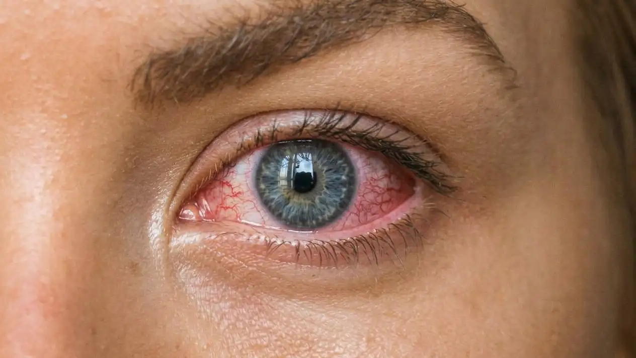
- A red eye.
- Strong discomfort with light.
- Blurred vision.
- Watering or discharge.
- A white or greyish spot on the cornea, sometimes visible in the mirror.
- A swollen eyelid and a feeling that something is in your eye.
If you wear contact lenses and your eye is red and painful, take them out and ask to be seen that same day. Do not cover the eye with a patch: it helps the bacteria. Bring your lenses, the case and the solution you use, because they can help identify the germ.
Risk factors
By far the biggest risk is wearing contact lenses, especially when they are not used as they should be:
- Sleeping in them, even for a nap or a single night. This is the mistake that raises the risk the most. The same goes for orthokeratology lenses, which are worn overnight to reshape the cornea.
- Water: showering, swimming in a pool or the sea, or getting into a hot tub with them in, or rinsing the lenses or the case with tap water.
- A dirty or old case, and topping up the solution instead of replacing it.
- Wearing lenses for longer than the manufacturer says, or handling them without washing your hands.
It can also happen without contact lenses. The risk goes up with:
- A knock or a scratch to the eye, especially from branches, soil or plant material.
- A very dry eye, eyelids that do not close properly or lashes that rub against the cornea.
- Previous surgery on the cornea or for cataracts.
- Long-term use of steroid eye drops or anaesthetic drops.
- Diabetes and a weakened immune system.
How it is diagnosed
With the clinic microscope (the slit lamp) I look at the size, depth and appearance of the lesion. A drop of an orange dye, fluorescein, shows up the area where the epithelium is missing. I also measure your vision, so we have a reference point.
Small ulcers away from the centre usually heal well with standard treatment. If the ulcer is large, central, deep or looks unusual, a sample is taken by gently scraping the surface with the eye numbed, in order to do a culture: this shows which germ it is and which antibiotic works against it. Sometimes the contact lens and the case are cultured too.
Not all corneal infections are caused by bacteria. Fungi, or a parasite called acanthamoeba, linked mainly to water and contact lenses, are treated differently.
Treatment
Antibiotic eye drops, and very often
The treatment is antibiotic drops. At first they are very frequent, often every hour, including at night for the first few days. It seems like a lot, but the cornea needs the antibiotic to be there all the time. Depending on the case, one or two types of drops are used, and for the most serious ulcers fortified drops are made up, which are more concentrated.
As the eye improves, they are spaced out. Do not stop them or cut them down on your own, even if it no longer hurts.
Steroids: never on your own
Sometimes, once the infection is under control, a steroid eye drop is added so that the scar is smaller. The ophthalmologist decides this, once the germ has been identified or the eye is responding well. Used too early, it can make the infection much worse.
If it does not respond
When the infection does not improve, the cultures are repeated and the treatment is adjusted. In severe cases where the cornea has become very thin, other measures may be needed, such as tissue glue, an amniotic membrane or a corneal transplant.
Before you put anything in your eye
Recovery and check-ups
The first check-ups are very close together, often daily or every other day. In the first 24 to 48 hours it is normal for the eye not to look any better, and with certain bacteria it may even be a little more inflamed. What counts is that the pain, the discharge and the spot get less after that.
When the infection heals, a scar may be left. If it is away from the centre, it hardly affects your sight. If it is in the centre, it can leave blurred or distorted vision, and sometimes a special contact lens or surgery helps to recover it.
- Stick to the times for your drops. An alarm on your phone helps, at night too.
- Do not put contact lenses back in until I tell you, and throw away the ones you were wearing and the case.
- If the pain gets worse or you see less well, call without waiting for your appointment.
Looking after your contact lenses
Contact lenses are safe if they are used properly. Almost all the infections linked to them come from the same habits:
- Wash and dry your hands well before touching them.
- Do not sleep in them, unless your ophthalmologist has specifically told you to.
- No water at all: not from the tap, the pool or the shower. Not for rinsing the case either.
- Always use fresh solution. Do not top up what was left in the case.
- Rub and rinse your lenses with their solution every time you take them out.
- Clean the case, leave it to dry upside down and replace it every three months at the most.
- Stick to the wearing time for each lens: daily, fortnightly or monthly.
- Coloured or novelty contact lenses are medical devices too. Only with a proper fitting and follow-up.
If you go back to wearing them after keratitis, it is worth reviewing the type of lens and how you use them. Single-use daily disposables cause fewer problems than reusable ones. At the clinic I carry out contact lens fitting and help you choose the one that suits you best.
Do you wear contact lenses and have a red, painful eye?
Take them out and call today. Do not wait to see if it goes away on its own.